
As noted by Spriggs and Boddington (1989), mesothelial cells often form papillary, pseudo-acinar structures in which the centre may appear excavated, filled with
homogeneous extracellular matrix. Such clusters are known as clusters with collagen cores (Delahaye et al, 1990). Both normal, reactive and
proliferative mesothelial cells may exhibit collagen production. Electron microscopy and immunohistochemistry have demonstrated that the collagen cores were composed of collagen type III
(reticulin) and some elastin embedded in extracellular matrix (mainly glycosaminoglycanes).
In patients with malignant mesothelioma, clusters with collagen cores may be demonstrated in as much as 50% of Giemsa stained specimens. Collagen cores seem to occur rarely in serous effusions
containing metastatic adenocarcinoma. They must not be confounded with mucus secretion. In mucus-secreting adenocarcinoma cells, intracellular mucin can be revealed by PAS or antibodies against
CEA, in contrast with mesothelial cells that do not have this capacity.
Key message for students in anatomic pathology
Do not confound collagen cores in normal, reactive and proliferative mesothelial cells with mucin-producing adenocarcinoma cells
Illustrations
Case provided by E. Piaton: 76-year-old woman. Bilateral annexectomy for suspicious left ovarian mass. The
peritoneal fluid (09EC04330) was considered as negative for tumour cells, but contained reactive mesothelial cells with collagen cores. Histopathology of the left ovarian mass (09EH04300): to be
completed

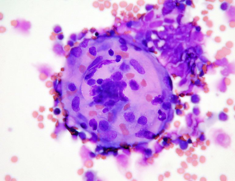
Figures 1-2: MGG stain x20 and x40
References
Delahaye M, De Jong AAW, Versnel MA, Hoosgsteden HC, Teeling P, Van der Kwast TH. Cytopathology of malignant mesothelioma. Reappraisal of the diagnostic value of
collagen cores. Cytopathology 1990, 1, 137-145
Spriggs AI, Boddington MM. Atlas of serous fluid cytopathology. A guide to the cells of pleural, pericardial, peritoneal and hydrocele fluids. Kluwer Academic Publishers, Dordrecht/Boston/London,
1989, p. 15


