
Provided by E. Piaton
Case report
A 70-year-old female patient with history of superficial bladder tumours (pTaG1-2) two years ago presented
post-menopausal bleeding. At colposcopy, an ulcerative and necrotizing cervical lesion was evidenced. Liquid-based cervical cytology (Thinprep 08EC04780) was interpreted as
"atypical glandular cells, suspicious for adenocarcinoma". according to the Bethesda system. Subsequent cervical biopsies showed typical papillary adenocarcinoma (08EH04606 - figs 1 and 2).
The patient underwent abdominal hysterectomy.
Six months later, she presented with a left supraclavicular lymph node which was sampled by microbiopsy under ultrasonography. Microscopic
examination revealed a metastatic serous papillary adenocarcinoma (08EH05204) comparable to that of the cervical site. The patient rapidly developed peritoneal carcinomatosis, and
microscopic examination of the peritoneal fluid (09EC04431 - figures 3 to 8) revealed numerous clusters and balls of adenocarcinomatous cells, papillary type. Typical small sized psammoma bodies (not shown) were evidenced in the centre of occasional papillary clusters.
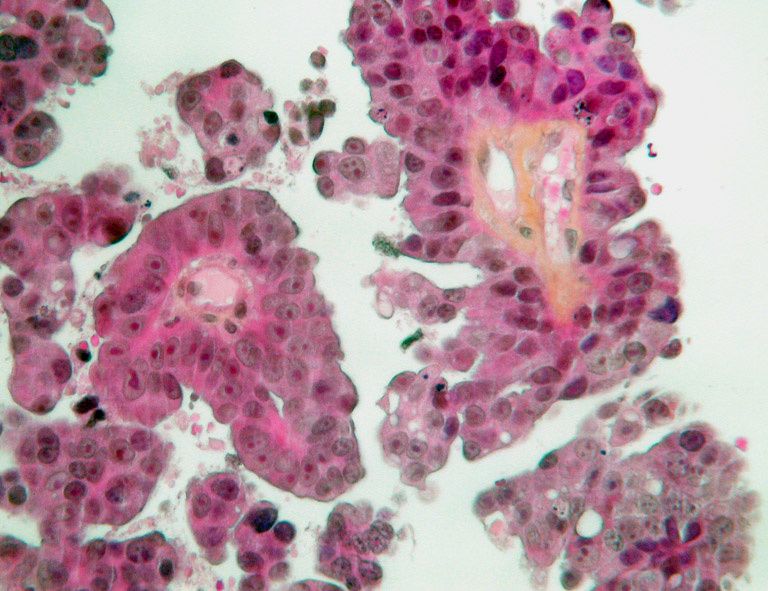

Figures 1 and 2: cervical biopsy (08EH04606). HES stain, x20 and
x40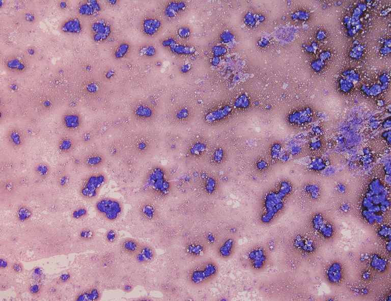
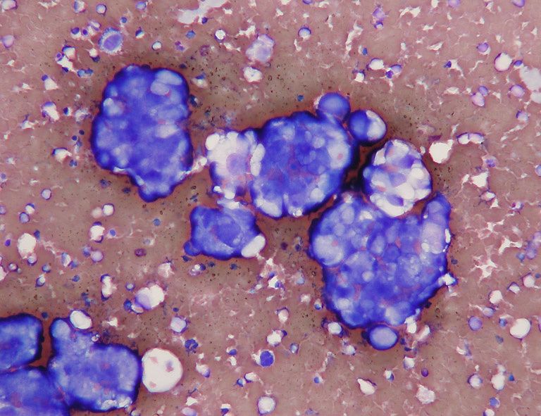
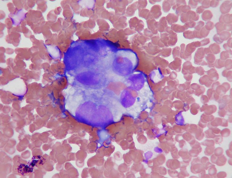
Figures 3-5: Peritoneal fluid (09EC04431). MGG stain, x4, x20 and x40 (centrifugation and smearing)
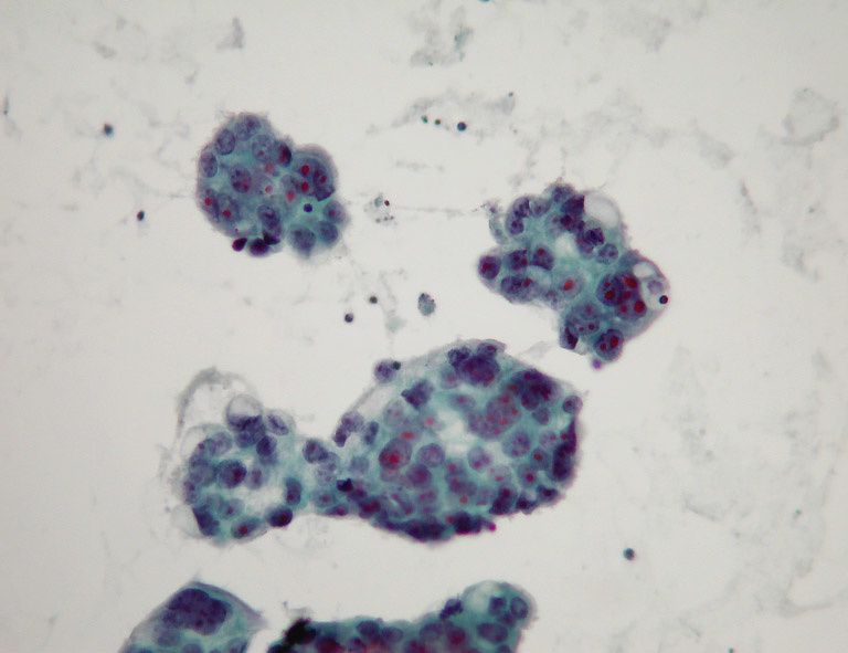
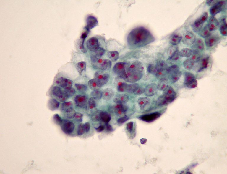
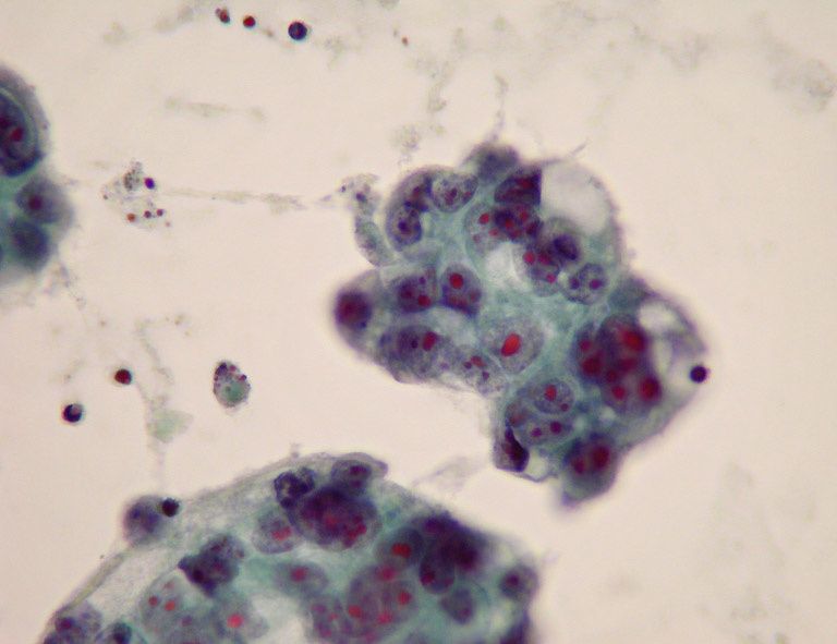
Figures 6-8: Peritoneal fluid (09EC04431). Papanicolaou stain, X20 and x40 (Thinprep* slides)


